Another RVOT VT: Retrograde 1:1 Conduction
Report:
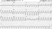
Ventricular tachycardia 168/min
Comment:
This is a good example where conduction sequences and QRS morphology do not help in making the diagnosis. Each QRS is followed after 0.18” by a sharp upright P wave in V1 consistent with a retrograde P wave; there are notches at the same position in the inferior leads but their polarity, as usual, may be difficult to determine20. The QRS is relatively short at 0.14” and its shape in V1 or V2 is a symmetric QS complex. This could be LBBB of supraventricular origin as much as ectopic VT.
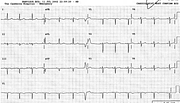
What helps is the right axis deviation over +100o in the frontal plane and a VEB practically identical to those in the paroxysm during sinus rhythm (Fig 16a). The latter is retrogradely conducted too, creating a longer than (fully) compensatory pause. This is, morphologically, another RVOT VT.
The compensatory pause is not always, as Schamroth put it, a “feeble reed to lean on”: in this case, despite the presence of sinus arrhythmia, one can make a valid inference of retrograde conduction from the resetting and post-ectopic depression of the SA node.
The left axis deviation ( a LAHB) in sinus rhythm is of no diagnostic significance vis à vis the VT. Similarly, the notching of sinus P waves may follow, transiently, either SVT or VT with retrograde conduction – the atria would not know the difference!
If you have any suggestions for or feedback on this report, please let us know.
Hi, can we chat about some terms and conditions?
The library and it's records are licensed under the Creative Commons Attribution 4.0 International license.
You are free to:
- Share — copy and redistribute the material in any medium or format for any purpose, even commercially.
- Adapt — remix, transform, and build upon the material for any purpose, even commercially.
- The licensor cannot revoke these freedoms as long as you follow the license terms.
Under the following terms:
- Attribution — You must give appropriate credit , provide a link to the license, and indicate if changes were made . You may do so in any reasonable manner, but not in any way that suggests the licensor endorses you or your use.
- No additional restrictions — You may not apply legal terms or technological measures that legally restrict others from doing anything the license permits.
By clicking agree below, you are agreeing to adhere to CC BY 4.0.

