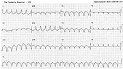
Arrhythmogenic Right Ventricular Dysplasia
Report:
Double sensitivity (1mV = 20mm)
Sinus rhythm 73/min
VEB
Right axis deviation (RAD) +110o
Right atrial abnormality (RAA)
Absolute small voltage (note the 20 mm/mV calibration)
Poor R wave progression
Nonspecific ST/T changes
Epsilon wave & V1/V6 duration c/c arrhythmogenic right ventricular dysplasia (ARVD)
Comment:
The patient presented originally with recurrent VT (Fig 88a) and eventually underwent extensive studies documenting her ARVD. The epsilon wave is the terminal “wrinkle” at the onset of the ST segment in lead V1; it corresponds to late small-amplitude potentials found at her EPS. It represents islands of late depolarisation often initiating reentry VT. The latter has a typical right ventricular morphology of LBBB (but with a slow S downslope in V1) and the (commonly found) LAD.
In ECGs taken at other times (not shown) there was T wave inversion in leads V1-3, also characteristic of ARVD. The trace shown has at least one other diagnostic criterion for ARVD: the QRS duration in V1 exceeds that of V6 by at least 0.06 sec33.
This patient also had an ICD which did not respond to the relatively slow VT shown below. It was programmed to fire at the rates over 160/min.
If you have any suggestions for or feedback on this report, please let us know.
Hi, can we chat about some terms and conditions?
The library and it's records are licensed under the Creative Commons Attribution 4.0 International license.
You are free to:
- Share — copy and redistribute the material in any medium or format for any purpose, even commercially.
- Adapt — remix, transform, and build upon the material for any purpose, even commercially.
- The licensor cannot revoke these freedoms as long as you follow the license terms.
Under the following terms:
- Attribution — You must give appropriate credit , provide a link to the license, and indicate if changes were made . You may do so in any reasonable manner, but not in any way that suggests the licensor endorses you or your use.
- No additional restrictions — You may not apply legal terms or technological measures that legally restrict others from doing anything the license permits.
By clicking agree below, you are agreeing to adhere to CC BY 4.0.

