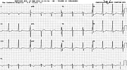
Right Axis Deviation in WPW Conduction
Report:
Sinus rhythm 88/min
Right axis deviation +110o
Wolff-Parkinson-White conduction, type ‘A’
Comment:
For ordinary clinical purposes WPW conduction is best divided into types ‘A’ and ‘B’, from Rosenbaum's now remote 1945 classification. Not only is it simple, it is easy to remember: in V1, ‘A’ is ‘above’ and ‘B’ is ‘below’ for the predominant QRS deflection. Type ‘A’ has the bypass bundle of Kent inserting into the left ventricle, type ‘B’ into the right.
Here, like in the previous Case 220 there is an illusion of normal PR interval. In V6, again, there appears to be a normal PR segment – but the simultaneous V4 and V5 show that δ wave starts from the end of the P wave.
The RAD here has no clinical significance, but can be, along with the dominant R in V1, mistaken for a sign of RVH.
Another example of RAD due to WPW conduction is shown below (Fig 21a).
The conduction becomes syndrome when there are attributable tachyarrhythmias, most commonly SVT, most dangerously AF.
If you have any suggestions for or feedback on this report, please let us know.
Hi, can we chat about some terms and conditions?
The library and it's records are licensed under the Creative Commons Attribution 4.0 International license.
You are free to:
- Share — copy and redistribute the material in any medium or format for any purpose, even commercially.
- Adapt — remix, transform, and build upon the material for any purpose, even commercially.
- The licensor cannot revoke these freedoms as long as you follow the license terms.
Under the following terms:
- Attribution — You must give appropriate credit , provide a link to the license, and indicate if changes were made . You may do so in any reasonable manner, but not in any way that suggests the licensor endorses you or your use.
- No additional restrictions — You may not apply legal terms or technological measures that legally restrict others from doing anything the license permits.
By clicking agree below, you are agreeing to adhere to CC BY 4.0.

