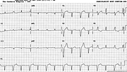
Alternating LBBB
Report:
Sinus rhythm 87/min
Alternating (2:1) left bundle branch block
Small frontal plane voltage
Nonspecific T wave changes
Comment:
The patient was admitted to CCU because of chest pain; a new LBBB developed. However, when disseminated carcinoma became obvious, he was placed on morphine infusion and allowed to die.
The tracing shows LBBB conduction in alternate beats. The PR interval is the same for both LBBB and normally conducted complexes: there is no bigeminy (diagnosed by the computer). There is no electrical alternans either: its definition requires not only fixed pacemaker but also fixed conduction. The malignancy, with a hint of tamponade, is just a red herring here. Of course, mechanical pulsus alternans could have been present, due to altered systolic mechanics in LBBB conduction. The only feature supporting pericardial involvement is the low voltage in limb leads but its significance – in this or any other context – remains uncertain.
The normally conducted beats show minor T wave changes, devoid of any diagnostic specificity.
An echocardiogram showed pericardial fluid but no evidence of tamponade.
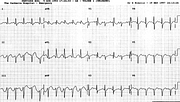
Alternation is common in ECGs. An alternating RBBB, with fixed LAHB, from a different patient, is shown below (Fig 31a).
If you have any suggestions for or feedback on this report, please let us know.
Hi, can we chat about some terms and conditions?
The library and it's records are licensed under the Creative Commons Attribution 4.0 International license.
You are free to:
- Share — copy and redistribute the material in any medium or format for any purpose, even commercially.
- Adapt — remix, transform, and build upon the material for any purpose, even commercially.
- The licensor cannot revoke these freedoms as long as you follow the license terms.
Under the following terms:
- Attribution — You must give appropriate credit , provide a link to the license, and indicate if changes were made . You may do so in any reasonable manner, but not in any way that suggests the licensor endorses you or your use.
- No additional restrictions — You may not apply legal terms or technological measures that legally restrict others from doing anything the license permits.
By clicking agree below, you are agreeing to adhere to CC BY 4.0.

