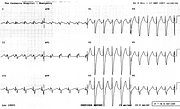
Large Right Precordial R Waves in LBBB
Report:
Sinus tachycardia 120/min
Left atrial abnormality (LAA)
Left bundle branch block
Probable old anterior infarction
Comment:
The right precordial are waves look typically thin, like antennæ or unipolar pacing spikes. There is also a loss of R amplitude in V4-5. This was not positional: other ECGs consistently showed this pattern (Fig 35a below). She had the same pattern while in incomplete LBBB one year earlier.
While suggestive of anterior infarction – even in the presence of LBBB17 – this pattern occurs, as in this patient, in primary congestive cardiomyopathy (COCM). She had marked 4-chamber enlargement and moderate mitral incompetence, with LV ejection fraction of only 20%.
Marked R wave notching in leads 1 and aVL also suggests previous infarction. It should be remembered that primary congestive cardiomyopathy may mimic infarction not just electrocardiographically, but also in showing segmental wall motion abnormalities or localised deficits in thallium uptake! This leaves coronary angiography and myocardial biopsy, but even these can be misleading at times. Autopsy is best, but by then it’s rather late.
If you have any suggestions for or feedback on this report, please let us know.
Hi, can we chat about some terms and conditions?
The library and it's records are licensed under the Creative Commons Attribution 4.0 International license.
You are free to:
- Share — copy and redistribute the material in any medium or format for any purpose, even commercially.
- Adapt — remix, transform, and build upon the material for any purpose, even commercially.
- The licensor cannot revoke these freedoms as long as you follow the license terms.
Under the following terms:
- Attribution — You must give appropriate credit , provide a link to the license, and indicate if changes were made . You may do so in any reasonable manner, but not in any way that suggests the licensor endorses you or your use.
- No additional restrictions — You may not apply legal terms or technological measures that legally restrict others from doing anything the license permits.
By clicking agree below, you are agreeing to adhere to CC BY 4.0.

