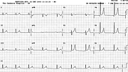
Marked Post-Thoracotomy ST Elevation
Report:
Sinus rhythm 77/min
Left ventricular hypertrophy voltage
ST segment elevation c/c pericarditis or ischæmia
Tall T waves ? ischæmic or hyperkalæmic
Comment:
What makes the trace suspicious is the combination of ST elevation and tall T waves. The elevation is rarely over 5 mm in pericarditis. Still, it is diffuse, without any reciprocal depression and the T waves could in fact be a normal variant.
The answer, as is often the case, is in the preoperative ECG (below, Fig 103a). The patient had a fairly typical early repolarisation pattern in the precordial leads, along with identical tall T waves and large voltage.
Knowing he had an aortic valve repair, volume overload LVH is a possibility, except that his valve was anatomically and functionally normal apart from an 8mm endothelial fibroelastoma on the left coronary cusp. Potassium, cardiac enzymes, etc were normal throughout.
Thus the explanation for the striking postoperative appearances is the combination of pre-existing early repolarisation and surgical pericarditis76.
If you have any suggestions for or feedback on this report, please let us know.
Hi, can we chat about some terms and conditions?
The library and it's records are licensed under the Creative Commons Attribution 4.0 International license.
You are free to:
- Share — copy and redistribute the material in any medium or format for any purpose, even commercially.
- Adapt — remix, transform, and build upon the material for any purpose, even commercially.
- The licensor cannot revoke these freedoms as long as you follow the license terms.
Under the following terms:
- Attribution — You must give appropriate credit , provide a link to the license, and indicate if changes were made . You may do so in any reasonable manner, but not in any way that suggests the licensor endorses you or your use.
- No additional restrictions — You may not apply legal terms or technological measures that legally restrict others from doing anything the license permits.
By clicking agree below, you are agreeing to adhere to CC BY 4.0.

