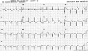
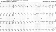
Anterior + Inferior MI
Report:
Sinus rhythm
Left atrial abnormality (LAA)
Borderline first degree AV block
Left axis deviation (LAD) – 60o
Left anterior hemiblock (LAHB)
Incomplete right bundle branch block (RBBB)
Extensive acute anterior myocardial infarction
Acute inferior myocardial infarction
Possible old posterolateral (1, aVL, R wave in V1) myocardial infarction
Left ventricular hypertrophy (LVH)
Comment:
Judging from the original angiogram report, the catastrophe was waiting to happen. The patient was too unwell to be asked why he did not have surgery; I speculated his insurance cover was not sufficient.
Leads 3 and aVF still show rS pattern of the (possibly pre-existing) LAHB; there are also R waves in V2-3. Over the ensuing four days, these were all replaced by q or Q waves (Fig 104a), while the original Q waves in 1 and aVL have disappeared. The LVH voltage has also gone.
In inferior MIs, S waves deeper than 5 mm usually signify an associated LAHB. It is not possible to diagnose the LAHB in the trace below, but it is probably still present; a VCG (not available at the Canberra Hospital) would be able to define it.
The broad primary R wave in V1, associated with Q waves of similar width in 1 and aVL, implies a previous posterolateral MI. ST segment elevation in V1 – despite the RBBB – may signify a right ventricular infarction in the presence of acute inferior MI; it may equally well be part of the obviously extensive anterior MI. ECG is not a very reliable guide to the topography of infarction. Paul Wood said, somewhere – I quoted this before - that it matters less where the infarction is than whether one had occurred in the first place.
If you have any suggestions for or feedback on this report, please let us know.
Hi, can we chat about some terms and conditions?
The library and it's records are licensed under the Creative Commons Attribution 4.0 International license.
You are free to:
- Share — copy and redistribute the material in any medium or format for any purpose, even commercially.
- Adapt — remix, transform, and build upon the material for any purpose, even commercially.
- The licensor cannot revoke these freedoms as long as you follow the license terms.
Under the following terms:
- Attribution — You must give appropriate credit , provide a link to the license, and indicate if changes were made . You may do so in any reasonable manner, but not in any way that suggests the licensor endorses you or your use.
- No additional restrictions — You may not apply legal terms or technological measures that legally restrict others from doing anything the license permits.
By clicking agree below, you are agreeing to adhere to CC BY 4.0.

