ST Depression Myocardial Infarction
Report:
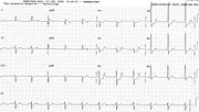
Sinus rhythm 97/min
Possible LVH
ST segment depression consistent with infarction/ischaemia
Comment:
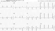
This is the worst ECG presentation for acute infarction – worse than T wave inversion or ST segment elevation77. The pattern is, in fact, that of main left coronary artery stenosis – diffuse depression with ST elevation in aVR > V1, as seen here. Infarction cannot be, of course, diagnosed on repolarisation changes alone – not without supporting laboratory and clinical data.
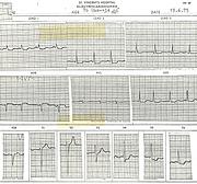
I arranged for this patient to return home to die, but it was not to be (Fig 106a, from my old ECG Library).
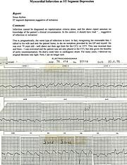
In Figs 106b and 106c , the extent of ST depression is more limited, but remained fixed over 72 hours, when the patient was sent to Sydney for possible surgery. The enzymes confirmed infarction, as in the case above. At angiography there were 90% stenoses in LAD, right and left circumflex arteries – more than the “main left” equivalent!
The ECG evolved day later to low-voltage LBBB seen here; it persisted until the patient’s death.
The reason I am no longer sure I was right in sending this patient home is that palliative care was not developed in those days and that, perhaps, the family prefers to see “everything done” before the final parting. Only if a clear-minded patient asked me himself not to admit him would I now send him back home.
Even counterpulsation would not be excluded now, when acute intervention is possible.
If you have any suggestions for or feedback on this report, please let us know.
Hi, can we chat about some terms and conditions?
The library and it's records are licensed under the Creative Commons Attribution 4.0 International license.
You are free to:
- Share — copy and redistribute the material in any medium or format for any purpose, even commercially.
- Adapt — remix, transform, and build upon the material for any purpose, even commercially.
- The licensor cannot revoke these freedoms as long as you follow the license terms.
Under the following terms:
- Attribution — You must give appropriate credit , provide a link to the license, and indicate if changes were made . You may do so in any reasonable manner, but not in any way that suggests the licensor endorses you or your use.
- No additional restrictions — You may not apply legal terms or technological measures that legally restrict others from doing anything the license permits.
By clicking agree below, you are agreeing to adhere to CC BY 4.0.


