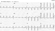
Non-Coronary Ischæmia
Report:
Sinus rhythm 80/min
Borderline first degree AV block
ST/T changes suggestive of ischæmia
Comment:
This is a good example of horizontal (“plane”) ST segment depression highly suggestive – almost diagnostic – of ischæmia. The associated T wave changes are more modest, but they, too, suggest ischæmia: TV1 > TV6 and T3 > T1. In this case, however, both are due to LVH engendered by severe aortic stenosis. The coronary arteries were normal and the LV function was impaired.
The LVH itself is not electrocardiographically visible: the repolarisation changes are atypical, while the voltages and the LAA are not there. Presumably the progressive LV dysfunction and myocardial damage are responsible, through a variety of mechanisms. Typical LVH with ST/T changes (and associated LAA) were present in earlier traces (Fig 15a).
The lesson is that one should try not to read too much into an ECG.
If you have any suggestions for or feedback on this report, please let us know.
Hi, can we chat about some terms and conditions?
The library and it's records are licensed under the Creative Commons Attribution 4.0 International license.
You are free to:
- Share — copy and redistribute the material in any medium or format for any purpose, even commercially.
- Adapt — remix, transform, and build upon the material for any purpose, even commercially.
- The licensor cannot revoke these freedoms as long as you follow the license terms.
Under the following terms:
- Attribution — You must give appropriate credit , provide a link to the license, and indicate if changes were made . You may do so in any reasonable manner, but not in any way that suggests the licensor endorses you or your use.
- No additional restrictions — You may not apply legal terms or technological measures that legally restrict others from doing anything the license permits.
By clicking agree below, you are agreeing to adhere to CC BY 4.0.

