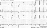
Acute Anterolateral Infarction
Report:
Sinus rhythm 68/min
VEBs
Acute anterolateral infarction
Left ventricular hypertrophy voltage
Comment:
The left circumflex artery was 100% blocked, but successfully dilated and stented at the PTCA. However, a sizeable posterolateral infarction remained. The posterior portion of the infarct (also seen scintigraphically) remains invisible on the ECG. Serum troponin rose to 157 µg/L (normal < 1.0 µ/L) and CPK to 2546 U/L (normal < 200 U/L). The LAD artery was diffusely diseased and the LV function globally depressed, with LVEF 41% at a subsequent radionuclide scan. The preserved large R waves in the anterolateral leads (below, day later) were somewhat misleading, presumably reflecting previously established LVH.
The reciprocal ST segment depression in V1-3 is indistinguishable from that produced by inferior infarctions; in this case it reflects a posterolateral one.
The two VEBs are good example of fully compensatory pause: the blocked sinus P waves can be seen as wrinkles in their ST segments. Below (Fig 34a), the VEBs become bigeminal and are seen in all the leads. They have overall LBBB morphology, but with slurred S descent in V1 and unusually rightward (about +85o) frontal axis.
If you have any suggestions for or feedback on this report, please let us know.
Hi, can we chat about some terms and conditions?
The library and it's records are licensed under the Creative Commons Attribution 4.0 International license.
You are free to:
- Share — copy and redistribute the material in any medium or format for any purpose, even commercially.
- Adapt — remix, transform, and build upon the material for any purpose, even commercially.
- The licensor cannot revoke these freedoms as long as you follow the license terms.
Under the following terms:
- Attribution — You must give appropriate credit , provide a link to the license, and indicate if changes were made . You may do so in any reasonable manner, but not in any way that suggests the licensor endorses you or your use.
- No additional restrictions — You may not apply legal terms or technological measures that legally restrict others from doing anything the license permits.
By clicking agree below, you are agreeing to adhere to CC BY 4.0.

