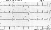
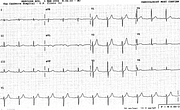
Small Ts in 1 and V6
Re-arrange ECGs to true time sequence, re-write report!
Report:
Sinus rhythm 59/min
T wave changes c/w ischæmia
Comment:
The TV1 > TV646 or T3 > T1 phenomenon is less well known than it should be. It is not normal, as most computer programmes would have it. The first set of cardiac injury markers was normal and the patient was lucky not to have been sent home. An (almost) normal trace belonging to this very patient prior to his current predicament is shown below (Fig 62a). At least the T waves were normal then; the anteroseptal ST segments tell a different story, soon to become more explicit. Wisely, he was admitted to CCU anyway, on account of typical pain.
What, unsurprisingly, happened two days later, is shown next (Fig 62b). This time the computer calls the acute anterolateral infarction “nonspecific lateral T wave changes”. It may be taking my own strategy of under-reporting too far.
He had CABGs 6 months later.
If you have any suggestions for or feedback on this report, please let us know.
Hi, can we chat about some terms and conditions?
The library and it's records are licensed under the Creative Commons Attribution 4.0 International license.
You are free to:
- Share — copy and redistribute the material in any medium or format for any purpose, even commercially.
- Adapt — remix, transform, and build upon the material for any purpose, even commercially.
- The licensor cannot revoke these freedoms as long as you follow the license terms.
Under the following terms:
- Attribution — You must give appropriate credit , provide a link to the license, and indicate if changes were made . You may do so in any reasonable manner, but not in any way that suggests the licensor endorses you or your use.
- No additional restrictions — You may not apply legal terms or technological measures that legally restrict others from doing anything the license permits.
By clicking agree below, you are agreeing to adhere to CC BY 4.0.


