Another Isolated U Wave Inversion
Report:
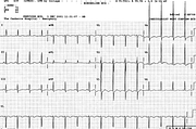
Sinus rhythm 95/min
Right atrial abnormality (RAA)
Probable LVH with ST/T changes
Inverted U waves c/w ischæmia
Movement artefact V5.
Comment:
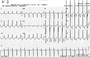
This patient, with chronic emphysema and hypertension, had an episode of chest pain two years previously and was admitted through Casualty with acute coronary insufficiency. The ECG at the time (Fig 72a) showed quite marked diffuse repolarisation changes well beyond what would be expected from LVH alone; these resolved over the following days. Thus, there is little doubt she had established and probably advanced coronary artery disease.
This time round, the evidence is subtler but no less serious. The negative U waves are best seen in V3 and V4, clearly later than the associated upright T waves in the same leads (and elsewhere). The gestalt here resembles Wellens’ syndrome, where it is the terminal T wave which is inverted. The clinical implications are the same.
For some reason, RAA is often seen in association with LVH – the so-called pseudo P pulmonale52. In this case, it is a real McCoy!
If you have any suggestions for or feedback on this report, please let us know.
Hi, can we chat about some terms and conditions?
The library and it's records are licensed under the Creative Commons Attribution 4.0 International license.
You are free to:
- Share — copy and redistribute the material in any medium or format for any purpose, even commercially.
- Adapt — remix, transform, and build upon the material for any purpose, even commercially.
- The licensor cannot revoke these freedoms as long as you follow the license terms.
Under the following terms:
- Attribution — You must give appropriate credit , provide a link to the license, and indicate if changes were made . You may do so in any reasonable manner, but not in any way that suggests the licensor endorses you or your use.
- No additional restrictions — You may not apply legal terms or technological measures that legally restrict others from doing anything the license permits.
By clicking agree below, you are agreeing to adhere to CC BY 4.0.

