Bigeminal AIVR: Inferoposterolateral MI
Report:
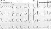
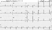
Sinus bradycardia (rate uncertain)
Accelerated idioventricular rhythm 77/min
Bigeminy ? exit block
Nonspecific ST/T changes
Comment:
The infarction cannot of course be diagnosed in the original tracing and what was left of its evidence after angioplasty is shown below (Fig 82a). Electrocardiographically, the evidence for inferior MI is strong, posterior MI reasonable and anterolateral one presumptive (with only T waves inverting). The overall gestalt is as stated in the title: inferoposterolateral MI. Paul Wood once said, somewhere, that it does not matter where the infarct is, but whether one had occurred.
The interesting feature is the AIVR, itself a marker of reperfusion. It shows a bigeminal pattern, presumably reflecting 3:2 conduction of the impulses from its focus to the ventricles. The focus may be somewhere in the left anterior-superior division of the left bundle branch since the QRS morphology resembles RBBB with LPHB. The rhythm slows in both the long and the short cycles and is followed by a sinus escape (rather than capture, as in the preceding two cases) beat. The last beat is again of the AIVR focus origin and comes rather quickly, with the same cycle length as at the beginning of the trace. The focus has “recovered”.
If you have any suggestions for or feedback on this report, please let us know.
Hi, can we chat about some terms and conditions?
The library and it's records are licensed under the Creative Commons Attribution 4.0 International license.
You are free to:
- Share — copy and redistribute the material in any medium or format for any purpose, even commercially.
- Adapt — remix, transform, and build upon the material for any purpose, even commercially.
- The licensor cannot revoke these freedoms as long as you follow the license terms.
Under the following terms:
- Attribution — You must give appropriate credit , provide a link to the license, and indicate if changes were made . You may do so in any reasonable manner, but not in any way that suggests the licensor endorses you or your use.
- No additional restrictions — You may not apply legal terms or technological measures that legally restrict others from doing anything the license permits.
By clicking agree below, you are agreeing to adhere to CC BY 4.0.

