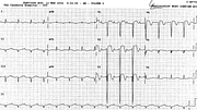
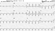
Anterior Infarction: “Trifascicular” Block
Report:
Sinus rhythm 99/min
First degree AV block
PR interval 0.28”
Right bundle branch block
Left anterior hemiblock
Axis –85o
Extensive acute anterior infarction
Comment:
The term “trifascicular” is a misnomer – irresistible to some – because the AV block is almost invariably situated not in a fascicle but within the AV node. The longer the PR interval, the more remote the likelihood of its “fascicular” provenance becomes.
Of course, a purist may have trouble diagnosing the LAHB itself59, especially if the LAD had been present earlier, associated with, or due to, inferior infarction (Fig 84a).
A feature of this tracing is the absence of reciprocal changes in the inferior leads despite impressive ST segment elevation in the anterior leads. This can be ascribed to (i) natural resolution of reciprocal changes over 20 hours since admission, (ii) new or increased LAHB, with deeper S waves in all three inferior leads and (iii) old inferior infarction, still ascertainable on the admission ECG (below, Fig 84a).
If you have any suggestions for or feedback on this report, please let us know.
Hi, can we chat about some terms and conditions?
The library and it's records are licensed under the Creative Commons Attribution 4.0 International license.
You are free to:
- Share — copy and redistribute the material in any medium or format for any purpose, even commercially.
- Adapt — remix, transform, and build upon the material for any purpose, even commercially.
- The licensor cannot revoke these freedoms as long as you follow the license terms.
Under the following terms:
- Attribution — You must give appropriate credit , provide a link to the license, and indicate if changes were made . You may do so in any reasonable manner, but not in any way that suggests the licensor endorses you or your use.
- No additional restrictions — You may not apply legal terms or technological measures that legally restrict others from doing anything the license permits.
By clicking agree below, you are agreeing to adhere to CC BY 4.0.

