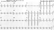
Ischæmic Giant T Wave Inversion
Report:
Sinus rhythm
Borderline first degree AV block
PR 0.22”
Incomplete left bundle branch block
QRS 0.12”
Deep T wave inversion consistent with infarction/ischæmia
Prolonged QTc 0.52”
Comment:
The patient had severe multivessel disease, with stenosed graft to the LAD dilated at PTCA the previous year; the left ventricle was normal. There was no explanation but ischæmia for her striking T wave inversion.
Neurogenic giant T wave inversion is, in fact, more asymmetrical and less pointed than this; the T wave is more splayed and the QT interval tends to be longer. Nevertheless, I would not (in fact, was not) sure that her ECG was not a marker of some cerebral event: I went to CCU to find out. Day later, the ECG showed more conventional pattern (106a). The same day, without further symptoms, her T waves normalised (Fig 106b).
A further anginal episode with ST segment depression and its resolution are shown in Figs 106c and 106d.
The conduction defect, reported as LBBB, could also be LAHB with added IVCD: the timing of secondary R wave in aVR with respect to aVL and the RS pattern in V6 support LAHB rather than LBBB. This does not matter much.
If you have any suggestions for or feedback on this report, please let us know.
Hi, can we chat about some terms and conditions?
The library and it's records are licensed under the Creative Commons Attribution 4.0 International license.
You are free to:
- Share — copy and redistribute the material in any medium or format for any purpose, even commercially.
- Adapt — remix, transform, and build upon the material for any purpose, even commercially.
- The licensor cannot revoke these freedoms as long as you follow the license terms.
Under the following terms:
- Attribution — You must give appropriate credit , provide a link to the license, and indicate if changes were made . You may do so in any reasonable manner, but not in any way that suggests the licensor endorses you or your use.
- No additional restrictions — You may not apply legal terms or technological measures that legally restrict others from doing anything the license permits.
By clicking agree below, you are agreeing to adhere to CC BY 4.0.

