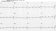
Hypocalcæmia
Report:
Sinus rhythm 70/min
Nonspecific lateral T wave changes
Prolonged QT interval
QTc 0.50”
Comment:
Although the QT interval is prolonged, the T wave is usually fairly normal in hypocalcæmia. This may explain the rarity of torsades de pointes: the repolarisation is delayed, but not dispersed. In this trace, there is a vertical heart position (aVL and aVF converge), making T3 > T1 a normal variant. However, this is more than that: T1 is not just smaller than T3, it is shallowly inverted. This may represent hypertensive or ischæmic heart disease.
If you have any suggestions for or feedback on this report, please let us know.
Hi, can we chat about some terms and conditions?
The library and it's records are licensed under the Creative Commons Attribution 4.0 International license.
You are free to:
- Share — copy and redistribute the material in any medium or format for any purpose, even commercially.
- Adapt — remix, transform, and build upon the material for any purpose, even commercially.
- The licensor cannot revoke these freedoms as long as you follow the license terms.
Under the following terms:
- Attribution — You must give appropriate credit , provide a link to the license, and indicate if changes were made . You may do so in any reasonable manner, but not in any way that suggests the licensor endorses you or your use.
- No additional restrictions — You may not apply legal terms or technological measures that legally restrict others from doing anything the license permits.
By clicking agree below, you are agreeing to adhere to CC BY 4.0.
