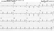
RVH in COAD
Report:
Atrial fibrillation, mean ventricular rate 85/min
Right axis deviation + 130o
qRV1, probable right ventricular hypertrophy
Nonspecific ST/T changes
Comment:
RVH is seldom expressed as dominant R wave in V1 in COAD; the commonest change is RAD in combination with persistent S waves in left precordial leads. The initial q wave in the qR complex seen here probably denotes right atrial enlargement92 (and is thus its only sign when P waves are absent).
The general sensitivity of ECG is low for RVH in COAD, less than 1/3 being clinically recognised. On the other hand, fully expressed RVH like this one is quite specific, and pulmonary hypertension can be safely predicted. This patient could be excluded from volume-reduction surgery on the basis of her ECG alone.
The QRS complex is characteristically thin in emphysema (0.07” here), although lower limit of normal QRS duration has not been defined in terms of any specific pathology.
The ventricular rate is controlled by drugs (digoxin and verapamil). There is a suggestion of sinus P waves in some cycles, but this is an illusion, recognised if one examines those phantom Ps in simultaneous channels.
An echocardiogram showed normal left and dilated right-sided chambers.
An ECG taken 5 years later is shown in Fig 124a below. She did rather well, considering.
If you have any suggestions for or feedback on this report, please let us know.
Hi, can we chat about some terms and conditions?
The library and it's records are licensed under the Creative Commons Attribution 4.0 International license.
You are free to:
- Share — copy and redistribute the material in any medium or format for any purpose, even commercially.
- Adapt — remix, transform, and build upon the material for any purpose, even commercially.
- The licensor cannot revoke these freedoms as long as you follow the license terms.
Under the following terms:
- Attribution — You must give appropriate credit , provide a link to the license, and indicate if changes were made . You may do so in any reasonable manner, but not in any way that suggests the licensor endorses you or your use.
- No additional restrictions — You may not apply legal terms or technological measures that legally restrict others from doing anything the license permits.
By clicking agree below, you are agreeing to adhere to CC BY 4.0.
