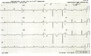
Cerebral T Waves
Report:
Sinus tachycardia 120/min
Vertical heart position
Left ventricular hypertrophy with atypical ST/T changes
Prolonged QT interval
Comment:
It is not possible to determine with certainty whether the QT interval is prolonged or U waves are present. The repolarisation changes look especially bizarre in the inferior leads.
They were quite impressive on admission to Casualty two days previously, but less bizarre (132a). Perhaps these represented the initial response to hypoxic stress from hanging.
The patient had very severe brain damage and died a few days later. The heart was normal at (Coroner’s) autopsy.
If you have any suggestions for or feedback on this report, please let us know.
Hi, can we chat about some terms and conditions?
The library and it's records are licensed under the Creative Commons Attribution 4.0 International license.
You are free to:
- Share — copy and redistribute the material in any medium or format for any purpose, even commercially.
- Adapt — remix, transform, and build upon the material for any purpose, even commercially.
- The licensor cannot revoke these freedoms as long as you follow the license terms.
Under the following terms:
- Attribution — You must give appropriate credit , provide a link to the license, and indicate if changes were made . You may do so in any reasonable manner, but not in any way that suggests the licensor endorses you or your use.
- No additional restrictions — You may not apply legal terms or technological measures that legally restrict others from doing anything the license permits.
By clicking agree below, you are agreeing to adhere to CC BY 4.0.


