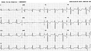
PR Segment Shift in Pericarditis
Report:
Sinus rhythm 88/min
PR segment shift consistent with pericarditis (best seen in leads 1, 2, aVR and V2)
Minimal ST elevation 2, 3, aVF
Comment:
The computer reported minimal ST segment elevation in the inferior leads, but I thought I knew better – it was the PR segment shift that made the ST segment look elevated. The T-P segment is the true baseline.
I was rather pleased to see shallow T wave inversion in her next ECG, consistent with pericarditis (133a). However, the PR segments were unchanged. I made the right diagnosis for the wrong reason!
By then I checked the patient’s record to make sure. She indeed had pericarditis, manifest as typical pain and a pericardial effusion. Dialysis & uræmic patients often fail to show any ECG signs of pericarditis. Note that the uræmic pericarditis is a different entity from the dialysis one97.
What, then, of those PR segments? Looking more closely, there is a borderline left atrial abnormality, best seen in the second P in V4 below. This is what done it!
If you have any suggestions for or feedback on this report, please let us know.
Hi, can we chat about some terms and conditions?
The library and it's records are licensed under the Creative Commons Attribution 4.0 International license.
You are free to:
- Share — copy and redistribute the material in any medium or format for any purpose, even commercially.
- Adapt — remix, transform, and build upon the material for any purpose, even commercially.
- The licensor cannot revoke these freedoms as long as you follow the license terms.
Under the following terms:
- Attribution — You must give appropriate credit , provide a link to the license, and indicate if changes were made . You may do so in any reasonable manner, but not in any way that suggests the licensor endorses you or your use.
- No additional restrictions — You may not apply legal terms or technological measures that legally restrict others from doing anything the license permits.
By clicking agree below, you are agreeing to adhere to CC BY 4.0.
