P Congenitale
Report:
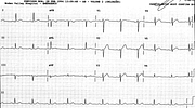
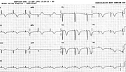
Sinus rhythm
First degree AV block
PR 0.22”
Right atrial abnormality
P height 5 mm in lead 2
P wave height > 1.5 mm in V1
P congenitale
P1 > P3 & P > 2.5 mm in a limb lead
Right axis deviation +230o
Probable right ventricular hypertrophy
Possible left ventricular hypertrophy
Comment:
The patient had, of course, biventricular hypertrophy; the ‘probable’ and the ‘possible’ above represent the constraints of electrocardiographic reporting. He had both an ASD and a VSD.
The P wave axis is only +40o due to large P in lead 1 and smaller one in lead 3. This is the pattern of P congenitale. Congenital heart disease productive of both right atrial hypertension and hypoxæmia produces the tallest P waves in cardiology. Mere distension from volume overload – e.g., in ASD – rarely produces very large P waves.
If you have any suggestions for or feedback on this report, please let us know.
Hi, can we chat about some terms and conditions?
The library and it's records are licensed under the Creative Commons Attribution 4.0 International license.
You are free to:
- Share — copy and redistribute the material in any medium or format for any purpose, even commercially.
- Adapt — remix, transform, and build upon the material for any purpose, even commercially.
- The licensor cannot revoke these freedoms as long as you follow the license terms.
Under the following terms:
- Attribution — You must give appropriate credit , provide a link to the license, and indicate if changes were made . You may do so in any reasonable manner, but not in any way that suggests the licensor endorses you or your use.
- No additional restrictions — You may not apply legal terms or technological measures that legally restrict others from doing anything the license permits.
By clicking agree below, you are agreeing to adhere to CC BY 4.0.


