S1S2S3 Pattern
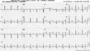
Report:
Sinus rhythm
Borderline biatrial abnormality
Indeterminate axis 200o
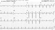
S1S2S3 pattern
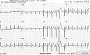
RSR’ V1
Comment:
The diagnosis of S1S2S3 pattern requires, strictly speaking, S wave larger than the corresponding R wave in the three standard leads, as in this trace. It is often seen in healthy individuals but, combined, as, again, in this trace, with RSR’ in V1 and prominent secondary R wave in that lead, it may be indistinguishable from RVH. The decision has to be made on other grounds.
As in emphysema with LAD, the S2 is deeper than S3, distinguishing it from LAHB, the commonest cause of LAD in adults.
The P waves are peaked but do not reach the 2.5 mm height required for the RAA; the PTF in V1 just makes 0.04”. Further curious feature is the notching of the R wave in leads 3 and aVF; it could pass for crochetage now recognised as a marker of ASD114. Some even find it a marker of patent foramen ovale (PFO) in ischæmic CVA115.
With all that, the patient’s only diagnoses were the psychiatric ones.
If you have any suggestions for or feedback on this report, please let us know.
Hi, can we chat about some terms and conditions?
The library and it's records are licensed under the Creative Commons Attribution 4.0 International license.
You are free to:
- Share — copy and redistribute the material in any medium or format for any purpose, even commercially.
- Adapt — remix, transform, and build upon the material for any purpose, even commercially.
- The licensor cannot revoke these freedoms as long as you follow the license terms.
Under the following terms:
- Attribution — You must give appropriate credit , provide a link to the license, and indicate if changes were made . You may do so in any reasonable manner, but not in any way that suggests the licensor endorses you or your use.
- No additional restrictions — You may not apply legal terms or technological measures that legally restrict others from doing anything the license permits.
By clicking agree below, you are agreeing to adhere to CC BY 4.0.


