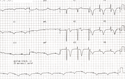
Post-LBBB T Wave Inversion
Report:
Sinus rhythm
Intermittent LBBB
Widespread symmetrical T wave inversion
Prolonged QT interval 0.60”
Comment:
This patient, with recurrent TIAs, could have post-syncopal T wave inversion. There was, however, no history of any “event” - cerebral or cardiac, and no clinical evidence for one. Most likely, the inversion is the result of previous LBBB conduction. It is also seen, at times, following periods of paced rhythm (also with LBBB morphology), VT or aberrant SVT.
The mechanism of T wave inversion following LBBB conduction is unknown132. The inversion is transient and its only significance lies in its differential diagnosis.
Below is an ECG entirely in normal conduction, but with a memory of what preceded it (Fig 175a).
If you have any suggestions for or feedback on this report, please let us know.
Hi, can we chat about some terms and conditions?
The library and it's records are licensed under the Creative Commons Attribution 4.0 International license.
You are free to:
- Share — copy and redistribute the material in any medium or format for any purpose, even commercially.
- Adapt — remix, transform, and build upon the material for any purpose, even commercially.
- The licensor cannot revoke these freedoms as long as you follow the license terms.
Under the following terms:
- Attribution — You must give appropriate credit , provide a link to the license, and indicate if changes were made . You may do so in any reasonable manner, but not in any way that suggests the licensor endorses you or your use.
- No additional restrictions — You may not apply legal terms or technological measures that legally restrict others from doing anything the license permits.
By clicking agree below, you are agreeing to adhere to CC BY 4.0.

