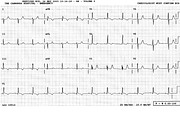
RVH
Report:
Sinus rhythm 70/min
Right axis deviation + 130o
qRV1 – right ventricular hypertrophy
Comment:
Most ECGs looking like this would suggest primary pulmonary hypertension, especially in a young woman. The heart has a limited repertoire: this example of RVH is the result of the RV being the systemic ventricle, due to congenital transposition (TGA) treated – now decades ago – by Mustard operation. The latter is, functionally, an atrial rather than an arterial switch.
The P waves are unremarkable – certainly no P pulmonale of emphysema – but do have a rightward axis, certainly over +70o.
Given the marked improvement in surgical treatment of congenital malformations30, there are now as many adult survivors as children with history of corrective or palliative surgery31.
If you have any suggestions for or feedback on this report, please let us know.
Hi, can we chat about some terms and conditions?
The library and it's records are licensed under the Creative Commons Attribution 4.0 International license.
You are free to:
- Share — copy and redistribute the material in any medium or format for any purpose, even commercially.
- Adapt — remix, transform, and build upon the material for any purpose, even commercially.
- The licensor cannot revoke these freedoms as long as you follow the license terms.
Under the following terms:
- Attribution — You must give appropriate credit , provide a link to the license, and indicate if changes were made . You may do so in any reasonable manner, but not in any way that suggests the licensor endorses you or your use.
- No additional restrictions — You may not apply legal terms or technological measures that legally restrict others from doing anything the license permits.
By clicking agree below, you are agreeing to adhere to CC BY 4.0.
