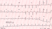
P Wave or T Wave?
Report:
Sinus bradycardia 37/min.
Left atrial abnormality .
First degree AV block.
Left bundle branch block
Comment:
The T wave is peaked and sharply demarcated from the preceding ST segment, mimicking a P' wave. Sequential strips (Fig 224a below) gradually dispel the illusion.
T wave is commonly notched in V2 or V3 in juvenile ECGs; here the angulation and the shape are probably due to progressive hyperkalæmia. The patien was no juvenile, either.
If you have any suggestions for or feedback on this report, please let us know.
Hi, can we chat about some terms and conditions?
The library and it's records are licensed under the Creative Commons Attribution 4.0 International license.
You are free to:
- Share — copy and redistribute the material in any medium or format for any purpose, even commercially.
- Adapt — remix, transform, and build upon the material for any purpose, even commercially.
- The licensor cannot revoke these freedoms as long as you follow the license terms.
Under the following terms:
- Attribution — You must give appropriate credit , provide a link to the license, and indicate if changes were made . You may do so in any reasonable manner, but not in any way that suggests the licensor endorses you or your use.
- No additional restrictions — You may not apply legal terms or technological measures that legally restrict others from doing anything the license permits.
By clicking agree below, you are agreeing to adhere to CC BY 4.0.
