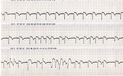
Agonal Junctional Tachycardia
Report:
Sinus bradycardia
Junctional tachycardia
AV dissociation
VEBs, bigeminy (below)
Second degree AV block, 2:1 (middle strip)
Third degree AV block, ventricular standstill
(Agonal) ST segment elevation
Comment:
The last part of a normal human heart to cease electrical activity is the right atrium, perhaps represented here by the positive P wave (initially triphasic) in the MCL1.
The run of bigeminy shown below (Fig 250a) occurred two minutes after the ventricular standstill recorded on the original strips. Agonal rhythms often recover electrical activity just as one thinks it’s all over. It may be of some clinical importance in that it may startle the assembled family whose eyes tend to become fixed on the monitor. The (presumptive) VEBs here look more normal than the supraventricular QRS complexes; such are the limitations of a single lead.
Shallow atrial repolarisation (Ta wave) is well shown in the bottom strip of the original recording.
If you have any suggestions for or feedback on this report, please let us know.
Hi, can we chat about some terms and conditions?
The library and it's records are licensed under the Creative Commons Attribution 4.0 International license.
You are free to:
- Share — copy and redistribute the material in any medium or format for any purpose, even commercially.
- Adapt — remix, transform, and build upon the material for any purpose, even commercially.
- The licensor cannot revoke these freedoms as long as you follow the license terms.
Under the following terms:
- Attribution — You must give appropriate credit , provide a link to the license, and indicate if changes were made . You may do so in any reasonable manner, but not in any way that suggests the licensor endorses you or your use.
- No additional restrictions — You may not apply legal terms or technological measures that legally restrict others from doing anything the license permits.
By clicking agree below, you are agreeing to adhere to CC BY 4.0.
