Acute Non-Embolic Cor Pulmonale
Report:
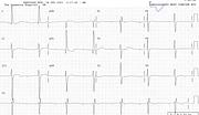
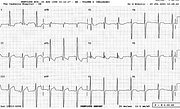
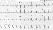
Sinus tachycardia 130/min
Normal axis
S1Q3T3 (McGinn-White) pattern consistent with acute cor pulmonale
RSR’ pattern V1
Precordial T wave inversion consistent with right ventricular strain
Comment:
The combined features are strongly suggestive of acute cor pulmonale.
In the context of pulmonary œdema (ARDS) in the single lung, the eponymous pattern did not add to the diagnosis of obvious and expected pulmonary hypertension. Yet, such is the power of words (S1Q3T3 pattern), that some members of the staff proposed “ruling out” pulmonary embolism on the strength of the S1Q3T3 pattern they knew to be associated with it!
I have never seen (or know anyone who has) a white lung due to pulmonary embolism. The contralateral one, perhaps (v. Case 203)! This patient of course did not have a contralateral lung.
If you have any suggestions for or feedback on this report, please let us know.
Hi, can we chat about some terms and conditions?
The library and it's records are licensed under the Creative Commons Attribution 4.0 International license.
You are free to:
- Share — copy and redistribute the material in any medium or format for any purpose, even commercially.
- Adapt — remix, transform, and build upon the material for any purpose, even commercially.
- The licensor cannot revoke these freedoms as long as you follow the license terms.
Under the following terms:
- Attribution — You must give appropriate credit , provide a link to the license, and indicate if changes were made . You may do so in any reasonable manner, but not in any way that suggests the licensor endorses you or your use.
- No additional restrictions — You may not apply legal terms or technological measures that legally restrict others from doing anything the license permits.
By clicking agree below, you are agreeing to adhere to CC BY 4.0.


