Idioventricular Rhythm Mime of RVH
Report:
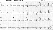
Idioventricular (?fascicular) rhythm 57/min
Giant T wave inversion
Prolonged QT interval
Comment:
The QRS morphology suggests, superficially, RVH. In V1, however, it is not a true qR complex – there is a small primary R wave as well: it’s an rsR’ complex. In view of the known preceding chronic LBBB, it probably originates from the distal, unblocked LBBB, maybe its posterior division (with LAHB axis in the frontal leads).
The T waves and their long QT interval attest to a preceding cerebral event, syncope or worse (actually the latter – she never woke up). This is consistent with arresting while waiting for a pacemaker.
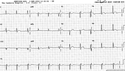
Below (Fig 59a) is her junctional rhythm the next day, looking like an incomplete LBBB with old anterior MI. The striking repolarisation changes have resolved.
If you have any suggestions for or feedback on this report, please let us know.
Hi, can we chat about some terms and conditions?
The library and it's records are licensed under the Creative Commons Attribution 4.0 International license.
You are free to:
- Share — copy and redistribute the material in any medium or format for any purpose, even commercially.
- Adapt — remix, transform, and build upon the material for any purpose, even commercially.
- The licensor cannot revoke these freedoms as long as you follow the license terms.
Under the following terms:
- Attribution — You must give appropriate credit , provide a link to the license, and indicate if changes were made . You may do so in any reasonable manner, but not in any way that suggests the licensor endorses you or your use.
- No additional restrictions — You may not apply legal terms or technological measures that legally restrict others from doing anything the license permits.
By clicking agree below, you are agreeing to adhere to CC BY 4.0.

