Emphysema: Left Axis Deviation
Report:
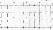
Sinus tachycardia
Right atrial abnormality
Left axis deviation –40o
Possible old anterior infarction
Suggestive of emphysema
Comment:
Characteristically, S2 > S3 in LAD due to emphysema vis à vis LAHB. Some argue this is not a true LAD but an “axis illusion” due to cardiac position and stretch in an overexpanded chest.
The precordial progression suggests an old anterior infarction, but there was no clinical evidence for this; emphysema alone can account for the QS complexes. She had incurable, crippling COAD and was extubated and allowed to die.
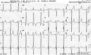
Below is a similar ECG (Fig 77a), from a 48 year old lady in similar circumstances. Her LAD could be from inferior MI, but wasn’t. There is small voltage in the frontal leads and poor R wave progression in the precordial ones. Like in the previous example, the QRS is typically narrow, although no actual criteria exist for abnormally short QRS. She also has a right ventricular impulse distorting ST segment in V3 (“electromechanical association”, v. Case 72).
If you have any suggestions for or feedback on this report, please let us know.
Hi, can we chat about some terms and conditions?
The library and it's records are licensed under the Creative Commons Attribution 4.0 International license.
You are free to:
- Share — copy and redistribute the material in any medium or format for any purpose, even commercially.
- Adapt — remix, transform, and build upon the material for any purpose, even commercially.
- The licensor cannot revoke these freedoms as long as you follow the license terms.
Under the following terms:
- Attribution — You must give appropriate credit , provide a link to the license, and indicate if changes were made . You may do so in any reasonable manner, but not in any way that suggests the licensor endorses you or your use.
- No additional restrictions — You may not apply legal terms or technological measures that legally restrict others from doing anything the license permits.
By clicking agree below, you are agreeing to adhere to CC BY 4.0.

