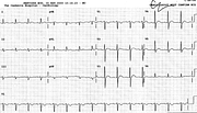
LVH – Volume Overload Pattern
Report:
Sinus rhythm 65/min
Left atrial abnormality
Left ventricular hypertrophy, volume overload pattern
RSR’ in V1-2
Comment:
The LAA is best seen in V3-5, along with prominent T waves. With LVH voltage in the chest leads this constitutes evidence for LV volume overload, from any cause. This patient had severe aortic and mild mitral incompetence.
The RSR’ pattern is not reflected in any S waves in 1 or V6: it’s unlikely to be an incomplete RBBB. Even if it were, it is a normal variant. So why report it? Just in case... One does not know the diagnosis reporting routine ECGs. She was only 51; there could have been an ASD, when RSR’ would be of significance.
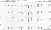
When, on another occasion, she was in heart failure precipitated by atrial fibrillation, the ECG evidence for LVH was not there (Fig 85a). Fluid accumulation and retention diminishes the voltages.
If you have any suggestions for or feedback on this report, please let us know.
Hi, can we chat about some terms and conditions?
The library and it's records are licensed under the Creative Commons Attribution 4.0 International license.
You are free to:
- Share — copy and redistribute the material in any medium or format for any purpose, even commercially.
- Adapt — remix, transform, and build upon the material for any purpose, even commercially.
- The licensor cannot revoke these freedoms as long as you follow the license terms.
Under the following terms:
- Attribution — You must give appropriate credit , provide a link to the license, and indicate if changes were made . You may do so in any reasonable manner, but not in any way that suggests the licensor endorses you or your use.
- No additional restrictions — You may not apply legal terms or technological measures that legally restrict others from doing anything the license permits.
By clicking agree below, you are agreeing to adhere to CC BY 4.0.

