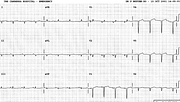
RVH in Emphysema
Report:
Sinus rhythm 96/min
Right axis deviation +170o
Right atrial abnormality
Right ventricular hypertrophy
Poor R wave progression
Comment:
The QRS axis is bizarrely right, with negative lead 2, and the emphysema QRS complex is characteristically narrow. In V1 it has a qR morphology, the q said to result from right atrial enlargement displacing the subjacent (to V1) right ventricle15.
The precordial R wave progression – or, rather, lack thereof – is due to emphysema, RVH and the marked frontal axis deviation; old anterior MI is a possibility, but an unlikely one in this setting.
The cor pulmonale is rarely so pronounced in ECGs. When it is, like here, it implies severe, advanced lung disease.
Below is another trace, taken a week earlier (Fig 9a). The voltages are lower, but the basic configuration is the same; P axis is still close to +90o. The computer reported LAA because of increased PTF in V1; this is almost certainly RAA in disguise.
If you have any suggestions for or feedback on this report, please let us know.
Hi, can we chat about some terms and conditions?
The library and it's records are licensed under the Creative Commons Attribution 4.0 International license.
You are free to:
- Share — copy and redistribute the material in any medium or format for any purpose, even commercially.
- Adapt — remix, transform, and build upon the material for any purpose, even commercially.
- The licensor cannot revoke these freedoms as long as you follow the license terms.
Under the following terms:
- Attribution — You must give appropriate credit , provide a link to the license, and indicate if changes were made . You may do so in any reasonable manner, but not in any way that suggests the licensor endorses you or your use.
- No additional restrictions — You may not apply legal terms or technological measures that legally restrict others from doing anything the license permits.
By clicking agree below, you are agreeing to adhere to CC BY 4.0.

