RBBB With a Difference
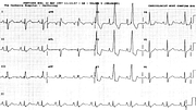
Report:
Sinus bradycardia
Borderline right axis deviation +90o
Right bundle branch block
V1 – V3 reversed
Comment:
The RBBB looks strikingly atypical in Lead V1, until one realises that V1 and V3 had been reversed! (Looking at the T wave helps).The trace was still somewhat unusual for an 18 year old, so I looked up his Casualty record. As often happens, it revealed little: pharyngitis, with “soft systolic murmur”. The latter must have been the reason ECG was taken. I was hoping for (at least) a fixed split of P2!
RBBB occurs with some frequency (much greater than that of LBBB) in “otherwise normal” hearts66.
Pharyngitis brings the now vanishing rheumatic fever to mind, but 1o AVB would be the expected finding, not RBBB. Also, too early, unless the pharyngitis is a recurrent one.
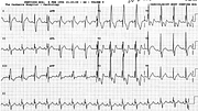
Oddly enough, when I had a routine ECG taken, the techician got flusterred and reversed the same leads (Fig 97a). I did not let him repeat the recording. I may use it some day, with its nice report.
If you have any suggestions for or feedback on this report, please let us know.
Hi, can we chat about some terms and conditions?
The library and it's records are licensed under the Creative Commons Attribution 4.0 International license.
You are free to:
- Share — copy and redistribute the material in any medium or format for any purpose, even commercially.
- Adapt — remix, transform, and build upon the material for any purpose, even commercially.
- The licensor cannot revoke these freedoms as long as you follow the license terms.
Under the following terms:
- Attribution — You must give appropriate credit , provide a link to the license, and indicate if changes were made . You may do so in any reasonable manner, but not in any way that suggests the licensor endorses you or your use.
- No additional restrictions — You may not apply legal terms or technological measures that legally restrict others from doing anything the license permits.
By clicking agree below, you are agreeing to adhere to CC BY 4.0.

