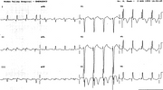
Atrial-Triggered Failed Pacemaker
Report:
Sinus rhythm 94 - 96/min 1
First degree AV block (PR 0.28") 2
Left atrial abnormality (LAA) 1
Atrial-triggered ventricular pacemaker 2
Failure to pace 3
LVH with ST/T changes (incomplete LBBB) ± ischæmia 1
Comment:
The pacing spikes bear constant relationship to the QRS complexes, but remain at a distance from them; they are in fact related to the preceding P waves. The ECG taken two days previously was unfortunately misreported (below), as the shorter PR interval placed - fortuitously - the pacing spike just before the onset of the QRS complex18. Ventricular capture by the pacemaker would have produced a broader QRS with left axis deviation à la LBBB. The observed axis would imply a superior displacement of the pacemaker tip toward the RV outflow tract.
The marked mid-precordial T wave inversion, especially the concordant one in V3, and in the absence of the corresponding ST segment depression associated with the LVH or incomplete LBBB, is indeed suspicious of ischaemia or other myocardial disease.
R complex in V6 is consistent with both septal infarction or (in this case incomplete) LBBB; both can also cause a QS in V1. A practical way of differentiating the two is by the size of the QRSs: septal infarcts diminish it, while incomplete LBBB increases it.
A great majority (90%) of patients with incomplete LBBB also have LVH.
If you have any suggestions for or feedback on this report, please let us know.
Hi, can we chat about some terms and conditions?
The library and it's records are licensed under the Creative Commons Attribution 4.0 International license.
You are free to:
- Share — copy and redistribute the material in any medium or format for any purpose, even commercially.
- Adapt — remix, transform, and build upon the material for any purpose, even commercially.
- The licensor cannot revoke these freedoms as long as you follow the license terms.
Under the following terms:
- Attribution — You must give appropriate credit , provide a link to the license, and indicate if changes were made . You may do so in any reasonable manner, but not in any way that suggests the licensor endorses you or your use.
- No additional restrictions — You may not apply legal terms or technological measures that legally restrict others from doing anything the license permits.
By clicking agree below, you are agreeing to adhere to CC BY 4.0.

