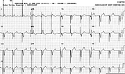
Negative Concordant Precordial Pattern
Report:
Sinus rhythm 1
Supraventricular ectopic beat 1
AV dissociation 5
Ventricular pacemaker rhythm 75/min with 100% capture 3
Comment:
At first it appears that the large (therefore unipolar) pacemaker spikes track the P waves, but the latter gradually merge into the QRS complexes (best seen in the rhythm strip) and disappear. This is not a “physiological” pacemaker (now usually a universal, or DDD, model).
Sinus P waves disappear after the 6th one, followed by a premature atrial wave; there is no definite atrial activity visible after that.
The paced rhythm has the expected pattern of LBBB with LAD, indicating right ventricular apical position of the pacemaker tip. The V leads are all negative. This constitutes a criterion of the ventricular origin of the rhythm, clearly not diagnostically important in this case (it would be if the spikes were absent). In VT, this is one of the criteria distinguishing it from aberrantly conducted SVT. It is not, however, as specific as its counterpart, the positive concordant precordial pattern.
Reverse pulsus paradoxus? Well, in AV dissociation the inspiration may put the P waves, fortuitously, just before the QRS complexes, bringing into play the atrial transport. It’s as simple as that. A distinction student should be able to adduce at least two further causes of reverse paradox36.
If you have any suggestions for or feedback on this report, please let us know.
Hi, can we chat about some terms and conditions?
The library and it's records are licensed under the Creative Commons Attribution 4.0 International license.
You are free to:
- Share — copy and redistribute the material in any medium or format for any purpose, even commercially.
- Adapt — remix, transform, and build upon the material for any purpose, even commercially.
- The licensor cannot revoke these freedoms as long as you follow the license terms.
Under the following terms:
- Attribution — You must give appropriate credit , provide a link to the license, and indicate if changes were made . You may do so in any reasonable manner, but not in any way that suggests the licensor endorses you or your use.
- No additional restrictions — You may not apply legal terms or technological measures that legally restrict others from doing anything the license permits.
By clicking agree below, you are agreeing to adhere to CC BY 4.0.
