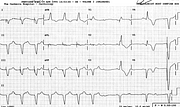
Failing Pacemaker
Report:
Sinus rhythm 1
Probable third degree AV block 1
AV dissociation 1 Ventricular escape beats 1
Primary T wave changes 1
Probable old anterior infarction 1
Pacemaker rhythm 72/min 1
Intermittent failure to pace 1
Probable lead displacement 2
Comment:
The bizarre frontal plane axis and the unexpected RBBB morphology of the paced beat in V1 indicate lead displacement, possibly a perforation. Failure to pace makes it even more likely. It could, however, be an epicardial pacemaker (it was not, historically).
The first escape beat is preceded by both a P wave and a pacing spike (best seen in V1), suggesting conduction of both, with ventricular fusion. The second escape beat, however, looks identical in the L2 rhythm strip at the bottom of the trace; its PR interval is longer and there is no spike artefact at the QRS onset. This means that the first beat is of the same (ectopic ventricular) provenance as the second.
In V6, the T wave of the escape beat is unexpectedly concordant with its QS complex, denoting a myocardial pathology supported by QR complexes in V4-5 above it. Even in ventricular ectopic beats, this indicates a previous infarction37.
If you have any suggestions for or feedback on this report, please let us know.
Hi, can we chat about some terms and conditions?
The library and it's records are licensed under the Creative Commons Attribution 4.0 International license.
You are free to:
- Share — copy and redistribute the material in any medium or format for any purpose, even commercially.
- Adapt — remix, transform, and build upon the material for any purpose, even commercially.
- The licensor cannot revoke these freedoms as long as you follow the license terms.
Under the following terms:
- Attribution — You must give appropriate credit , provide a link to the license, and indicate if changes were made . You may do so in any reasonable manner, but not in any way that suggests the licensor endorses you or your use.
- No additional restrictions — You may not apply legal terms or technological measures that legally restrict others from doing anything the license permits.
By clicking agree below, you are agreeing to adhere to CC BY 4.0.
