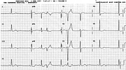
Mixed Bigeminy
Report:
Sinus rhythm 74/min
SVEBs, blocked
VEBs
Left ventricular hypertrophy with ST/T changes
Comment:
The diagnosis of sinus bradycardia is refuted by the negative (in the inferior leads) P waves – probably of junctional origin – after the first, fifth and sixth sinus beat. Marriott’s “commonest causes of pauses”135 follow the preceding QRSs by approximately 0.52”.
From the third sinus beat onward the rhythm is, in a sense, bigeminal: at first with the two VEBs and then with blocked SVEBs. The pulse would, most likely, reveal only a regular bradycardia 37/min.
In V1, the broad complex takes almost 0.08” to descend to its nadir: this makes a ventricular origin more likely than a supraventricular one, with LBBB aberrancy.
If the junctional extrasystoles were blocked retrogradely as well, the only evidence of their existence would be unexplained blocked P waves – a pseudoblock. Unfortunately, no examples could be found in this patient’s record. In the company of VEBs, the junctional SVEBs may well be main-stem (bundle of His) extrasystoles, with antegrade block. Standard ECG cannot tell them apart.
If you have any suggestions for or feedback on this report, please let us know.
Hi, can we chat about some terms and conditions?
The library and it's records are licensed under the Creative Commons Attribution 4.0 International license.
You are free to:
- Share — copy and redistribute the material in any medium or format for any purpose, even commercially.
- Adapt — remix, transform, and build upon the material for any purpose, even commercially.
- The licensor cannot revoke these freedoms as long as you follow the license terms.
Under the following terms:
- Attribution — You must give appropriate credit , provide a link to the license, and indicate if changes were made . You may do so in any reasonable manner, but not in any way that suggests the licensor endorses you or your use.
- No additional restrictions — You may not apply legal terms or technological measures that legally restrict others from doing anything the license permits.
By clicking agree below, you are agreeing to adhere to CC BY 4.0.
